44 light microscope with labels
LAS X Industry Microscope software for Industry | Products The software can handle multiple users who have different levels of microscope skills and diverse tasks to accomplish. Profiles according to user’s skills. The LAS X software enables you to create profiles according to the skills and tasks of individual users – from microscopy beginner to expert. It helps you to get reliable results. Microscope Objective Lens | Products | Leica Microsystems The objective lens is a critical part of the microscope optics. The microscope objective is positioned near the sample, specimen, or object being observed. It has a very important role in imaging, as it forms the first magnified image of the sample. The numerical aperture (NA) of the objective indicates its ability to gather light and largely determines the microscope’s …
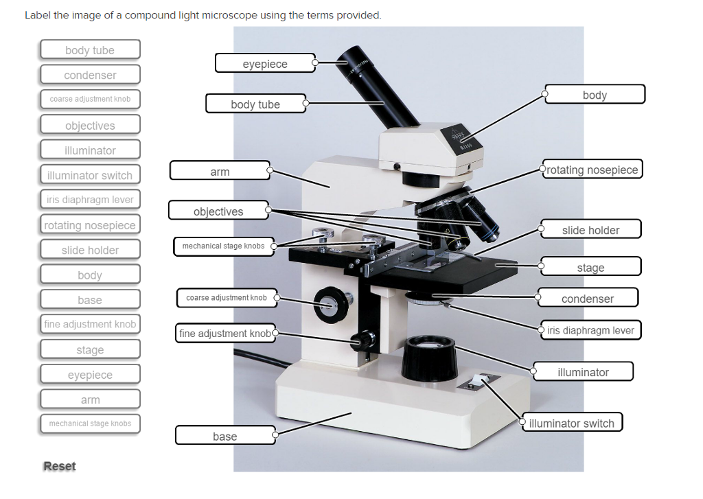
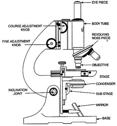
A Study of the Microscope and its Functions With a Labeled Diagram ... The microscope is an important instrument in the world of biological science. Diagrams have always been of great help in understanding both the structural and functional aspects of entities. These labeled microscope diagrams and the functions of its various parts, attempt to simplify the microscope for you.

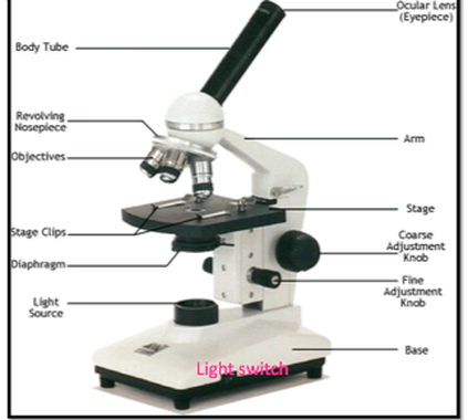
Light microscope with labels
Parts of the Microscope with Labeling (also Free Printouts) Parts of the Microscope with Labeling (also Free Printouts) By Editorial Team March 7, 2022 A microscope is one of the invaluable tools in the laboratory setting. It is used to observe things that cannot be seen by the naked eye. Table of Contents 1. Eyepiece 2. Body tube/Head 3. Turret/Nose piece 4. Objective lenses 5. Knobs (fine and coarse) 6. Microscope, Microscope Parts, Labeled Diagram, and Functions Majority of high quality microscopes used in laboratory include an Abbe condenser with an iris diaphragm. When iris diaphragm is combined with Abbe condenser, it control both the quantity of light applied as well as focus on the specimen. Aperture: It is the hole in the stage through which the base (transmitted) light reaches the stage. Parts of Stereo Microscope (Dissecting microscope) – labeled … A stereo microscope allows you to see the surface of specimens with a 3-dimensional view. Under a stereo microscope, you can see the metallic texture and colors of the mosquito’s compound eyes. In contrast, the light has to pass through the specimen to form the image under a compound microscope.
Light microscope with labels. Compound Microscope: Definition, Diagram, Parts, Uses, Working ... - BYJUS A compound microscope is defined as. A microscope with a high resolution and uses two sets of lenses providing a 2-dimensional image of the sample. The term compound refers to the usage of more than one lens in the microscope. Also, the compound microscope is one of the types of optical microscopes. The other type of optical microscope is a ... Microscope Diagram and Functions - Pinterest Download Clker's Microscope With Labels clip art and related images now. ... A diagram showing all of the parts of a compound light microscope. Microscope labels Flashcards | Quizlet Microscope label Learn with flashcards, games, and more — for free. Parts of a microscope with functions and labeled diagram Microscopic illuminator - This is the microscopes light source, located at the base. It is used instead of a mirror. It captures light from an external source of a low voltage of about 100v. Condenser - These are lenses that are used to collect and focus light from the illuminator into the specimen.
ZEISS Axioscope 5 Smart Laboratory Microscope Focus. Snap. Done. Forget about the 15 steps and clicks to document samples with multiple fluorescent labels. With Smart Microscopy, this is a thing of the past. Axioscope 5 with Axiocam 202 mono and Colibri 3 LED illumination take this workload from you. You keep your hands at the microscope stand. Relaxed. Compound Light Microscope: Everything You Need to Know A fluorescence microscope, also called a confocal microscope, is a kind of biological microscope that operates by using different light colors and wavelengths over-dyed specimen samples in order for the dye to interact with the light, after which the resulting image is scanned. Microscope Parts and Functions First, the purpose of a microscope is to magnify a small object or to magnify the fine details of a larger object in order to examine minute specimens that cannot be seen by the naked eye. Here are the important compound microscope parts... Eyepiece: The lens the viewer looks through to see the specimen. Microscope With Labels clip art - Pinterest Jul 3, 2012 - Download Clker's Microscope With Labels clip art and related images now ... A diagram showing all of the parts of a compound light microscope.
What is label in microscope? - Gowanusballroom.com Parts of the Microscope with Labeling (also Free Printouts) 1. Eyepiece. Through the eyepiece, you can visualize the object being studied. Its magnification capacity ranges between… 2. Body tube/Head. It is the structure that connects the eyepiece to the lenses. Image 2: The body tube part of a… Is there a print version of the microscope layout? Microscope Objective Lens | Products | Leica Microsystems The objective lens is a critical part of the microscope optics. The microscope objective is positioned near the sample, specimen, or object being observed. It has a very important role in imaging, as it forms the first magnified image of the sample. The numerical aperture (NA) of the objective indicates its ability to gather light and largely determines the microscope’s resolution, the ... Labeling the Parts of the Microscope Labeling the Parts of the Microscope This activity has been designed for use in homes and schools. Each microscope layout (both blank and the version with answers) are available as PDF downloads. You can view a more in-depth review of each part of the microscope here. Download the Label the Parts of the Microscope PDF printable version here. What is Electron Microscopy? - UMASS Medical School It is termed a scanning electron microscope because the image is formed by scanning a focused electron beam onto the surface of the specimen in a raster pattern. The interaction of the primary electron beam with the atoms near the surface causes the emission of particles at each point in the raster (e.g., low energy secondary electrons, high energy back scatter electrons, X-rays …

How does a microscope work? - Explain that Stuff
Microscope Labeling - The Biology Corner 1) Start with scanning (the shortest objective) and only use the COARSE knob . Once it is focused… 2) Switch to low power (medium) and only use the COARSE knob . You may need to recenter your slide. Once it is focused.. 3) Switch to high power (long objective).

Overview of a Light Microscope | Brewlab
Microscope Types (with labeled diagrams) and Functions This is an advanced microscope that has specific application in viewing, observing and measuring the optical thickness and phase of completely transparent specimens and objects. A tiny interferometer is used and a specimen is placed on beam path of it. This path is split and then rejoined to create two superimposed images of the specimen in focus.
Solved: Label The Image Of A Compound Light Microscope Usi... | Chegg.com
Compound Microscope Parts, Functions, and Labeled Diagram Compound Microscope Definitions for Labels. Eyepiece (ocular lens) with or without Pointer: The part that is looked through at the top of the compound microscope. Eyepieces typically have a magnification between 5x & 30x. Monocular or Binocular Head: Structural support that holds & connects the eyepieces to the objective lenses.
Microscope and its types |readbiology.com
Microscope Labeling Game - PurposeGames.com This is an online quiz called Microscope Labeling Game There is a printable worksheet available for download here so you can take the quiz with pen and paper. This quiz has tags. Click on the tags below to find other quizzes on the same subject. Science microsope Total Points 0 Get started! Today's Rank -- 0 's Points Points 15

7th Grade Science 2015-2016: History of and Using a Compound Microscope
Label the microscope — Science Learning Hub All microscopes share features in common. In this interactive, you can label the different parts of a microscope. Use this with the Microscope parts activity to help students identify and label the main parts of a microscope and then describe their functions. Drag and drop the text labels onto the microscope diagram.
Labeling A Compound Light Microscope - ClipArt Best
Solved Label the image of a compound light microscope using - Chegg Expert Answer. 100% (17 ratings) Transcribed image text: Label the image of a compound light microscope using the terms provided.
Topic 1.2 Ultra-Structure of Cells - AMAZING WORLD OF SCIENCE WITH MR. GREEN
Light Microscope- Definition, Principle, Types, Parts, Labeled Diagram ... A light microscope is a biology laboratory instrument or tool, that uses visible light to detect and magnify very small objects and enlarge them. They use lenses to focus light on the specimen, magnifying it thus producing an image. The specimen is normally placed close to the microscopic lens.

Chapter 16, Page 2 - HistologyOLM 4.0
ZEISS Lightsheet 7 – Light Sheet Microscope This effect occurs in all fluorescence microscopes, but the illumination axis in light sheet fluorescence microscopy is perpendicular to the observation axis and so this effect is more obvious. In Lightsheet 7, a patented Pivot Scanner alters the angle of the light sheet upwards and downwards during image acquisition.
Label Light Microscope - ClipArt Best
Electron microscope - Wikipedia An electron microscope is a microscope that uses a beam of accelerated electrons as a source of illumination. As the wavelength of an electron can be up to 100,000 times shorter than that of visible light photons, electron microscopes have a higher resolving power than light microscopes and can reveal the structure of smaller objects.. Electron microscopes use shaped magnetic …
Using a Light Microscope - AyushiSinhaMicroscopy
ZEISS Axioscope 5 Smart Laboratory Microscope Focus. Snap. Done. Forget about the 15 steps and clicks to document samples with multiple fluorescent labels. With Smart Microscopy, this is a thing of the past. Axioscope 5 with Axiocam 202 mono and Colibri 3 LED illumination take this workload from you. You keep your hands at the microscope stand. Relaxed.

Euglena Acus 2 - BF microscope 1250x - YouTube
Labeling the Parts of the Microscope - Pinterest Parts of Microscope with their Functions and Working Principle Optical Microscope, Microscope Parts, Electron. Parts of a Microscope and Their Functions.
Using the Microscope
Microscope Parts, Function, & Labeled Diagram - slidingmotion Condenser. The condenser is to focus the light, which passes from the microscopic illuminator to the specimen. This condenser is located just below the diaphragm. This diaphragm is one of the important parts of the compound microscope which will help to get an accurate and sharp image. The condenser has a magnification power of 400X and above.
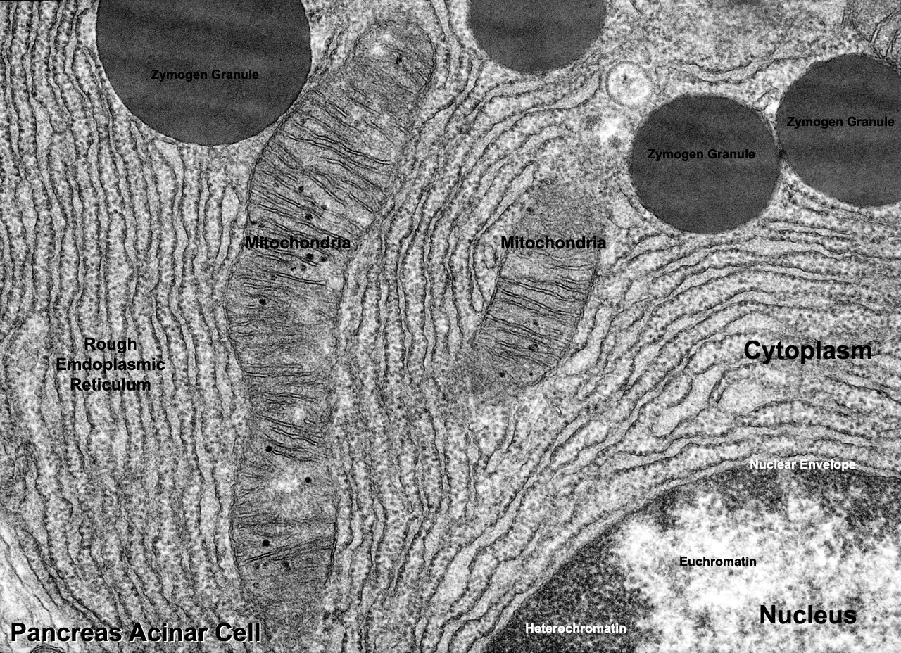
File:Pancreas acinar cell em01.jpg - Embryology
Electron microscope - Wikipedia An electron microscope is a microscope that uses a beam of accelerated electrons as a source of illumination. As the wavelength of an electron can be up to 100,000 times shorter than that of visible light photons, electron microscopes have a higher resolving power than light microscopes and can reveal the structure of smaller objects.
- Labeled Microscope - Virtual Fluorescent Microscope - Wartburg College Biology Department
Label the microscope — Science Learning Hub 08.06.2018 · All microscopes share features in common. In this interactive, you can label the different parts of a microscope. Use this with the Microscope parts activity to help students identify and label the main parts of a microscope and then describe their functions.. Drag and drop the text labels onto the microscope diagram. If you want to redo an answer, click on the …

OMAX Microscope 50mm Microscope Substage Mirror with 5mm Diameter Pin
Light Microscope: Functions, Parts and How to Use It To use a light microscope, you can follow the steps below carefully. Start with a low lens and a clean slide. The microscope stage should be lowered as low as possible. Center the slide so that the specimen is under the objective lens. Use the coarse adjustment knob to get a general focus. Then slowly move up the stage until focus is achieved.

Daphnia Heartbeat - YouTube
Light microscopes - Cell structure - Edexcel - BBC Bitesize Microscopes are used to produce magnified images. There are two main types of microscope: light microscopes are used to study living cells and for regular use when relatively low magnification and...
Search in gallery
Microscope, Anatomy bones, Diagram chart - Pinterest Light microscope, optical microscope diagrams. Label ... label microscope diagram | Charts Optical Microscope, Microscope Parts, Electron Microscope, ...
Post a Comment for "44 light microscope with labels"