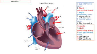
41 heart structure and labels
Heart: Anatomy and Function - Cleveland Clinic What are the parts of the heart's anatomy? The parts of your heart are like the parts of a house. Your heart has: Walls. Chambers (rooms). Valves (doors). Blood vessels (plumbing). Electrical conduction system (electricity). Heart walls Your heart walls are the muscles that contract (squeeze) and relax to send blood throughout your body. How to Draw the Internal Structure of the Heart (with Pictures) To draw the internal structure of a human heart, follow the steps below. Part 1 Finding a Diagram 1 To find a good diagram, go to Google Images, and type in "The Internal Structure of the Human Heart". Find an image that displays the entire heart, and click on it to enlarge it. 2 Find a piece of paper and something to draw with.
Heart Labels - Printable or Custom Printed Stickers | Avery.com Heart Labels Order blank or custom printed heart labels that you can personalize for product branding, promotions, wedding favors, party favors & any item that needs a little love. Blank Heart Labels Design & print personalized heart labels on-demand when you need them

Heart structure and labels
Heart anatomy: Structure, valves, coronary vessels | Kenhub The heart is shaped as a quadrangular pyramid, and orientated as if the pyramid has fallen onto one of its sides so that its base faces the posterior thoracic wall, and its apex is pointed toward the anterior thoracic wall. Human Heart - Diagram and Anatomy of the Heart - Innerbody Because the heart points to the left, about 2/3 of the heart's mass is found on the left side of the body and the other 1/3 is on the right. Anatomy of the Heart Pericardium. The heart sits within a fluid-filled cavity called the pericardial cavity. The walls and lining of the pericardial cavity are a special membrane known as the pericardium. Human Heart - Anatomy, Functions and Facts about Heart Following are the main functions of the heart: One of the primary functions of the human heart is to pump blood throughout the body. Blood delivers oxygen, hormones, glucose and other components to various parts of the body, including the human heart. The heart also ensures that adequate blood pressure is maintained in the body.
Heart structure and labels. Heart: illustrated anatomy - e-Anatomy - IMAIOS This interactive atlas of human heart anatomy is based on medical illustrations and cadaver photography. The user can show or hide the anatomical labels which provide a useful tool to create illustrations perfectly adapted for teaching. Anatomy of the heart: anatomical illustrations and structures, 3D model and photographs of dissection. Heart Anatomy Labeling Game - PurposeGames.com About this Quiz This is an online quiz called Heart Anatomy Labeling Game There is a printable worksheet available for download here so you can take the quiz with pen and paper. Your Skills & Rank Total Points 0 Get started! Today's Rank -- 0 Today 's Points One of us! Game Points 19 You need to get 100% to score the 19 points available Actions The structure of the heart - Structure and function of the heart ... The structure of the heart. If you clench your hand into a fist, this is approximately the same size as your heart. It is located in the middle of the chest and slightly towards the left. PDF Free Anatomy Coloring Page - North Carolina State University The ate.2S the heart With oxygen ate labeled with at'l Color these areas The areas o' the heart with less oxygen ate labeled with a color areas BLUE. ARTERY LEFT LUNG PULMONARY VEINS AORTA PULMONARY VEINS raGHT LUNG ATRIUM RIGHT VENTRICLE INFERIOR VFNACAVA LEFT LEFT VENTRICLE AORTA BODY Downloaded from azcoloring.com
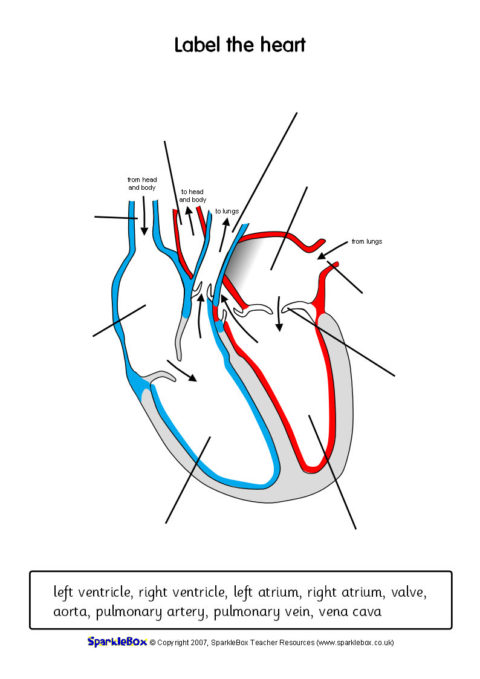
25 Blank Heart Diagram to Label | Softball Wristband Template May 16, 2020 - Blank Heart Diagram to Label - 25 Blank Heart Diagram to Label , Labled Diagram Word Wiring Diagrams. Pinterest. Today. Explore. When autocomplete results are available use up and down arrows to review and enter to select. Touch device users, explore by touch or with swipe gestures. Diagrams, quizzes and worksheets of the heart | Kenhub Worksheet showing unlabelled heart diagrams. Using our unlabeled heart diagrams, you can challenge yourself to identify the individual parts of the heart as indicated by the arrows and fill-in-the-blank spaces. This exercise will help you to identify your weak spots, so you'll know which heart structures you need to spend more time studying ... Heart Blood Flow | Simple Anatomy Diagram, Cardiac Circulation ... - EZmed The easiest way to understand the blood flow through the heart is to divide the heart into 2 sides. We first have the right side of the heart shown in blue below. There are 6 main steps or structures in which blood flows through the right side of the heart. Next, we have the left side of the heart shown in red. Human Heart (Anatomy): Diagram, Function, Chambers, Location ... Human Heart (Anatomy): Diagram, Function, Chambers, Location in Body The right atrium receives blood from the veins and pumps it to the right ventricle. The right ventricle receives blood from the...
A Diagram of the Heart and Its Functioning Explained in Detail Human heart is covered by a double layered structure which is known as pericardium. The outer layer is associated with the major blood vessels whereas the inner layer is attached to the cardiac muscles. These layers are separated by a pericardial fluid. This covering is like a membrane which holds all the parts of the heart. Chambers Heart Diagram with Labels and Detailed Explanation The heart is located under the ribcage, between the lungs and above the diaphragm. It weighs about 10.5 ounces and is cone shaped in structure. It consists of the following parts: Heart Detailed Diagram Heart - Chambers There are four chambers of the heart . The upper two chambers are the auricles and the lower two are called ventricles. heart diagram and labels Heart diagram label labels human without. Anatomy of heart interior with labels — cross section, blood vessels. The Heart: Structure and Function - YouTube. 11 Pictures about The Heart: Structure and Function - YouTube : File:Heart diagram-en.svg - Wikipedia, Heart Diagram To Label - ClipArt Best and also Label the Heart Worksheets (SB6634 ... Human Heart Diagram Labeled | Science Trends Jan 01, 2019 · List Of Heart Structures Heart Chambers Ventricles – The bottom two heart chambers. Atra – The upper two heart chambers. Wall Of The Heart Sinoatrial Node – A collection of tissue that releases electrical impulses and defines the rate of contraction for the heart. Atrioventricular Bundle – The fibers which transmit cardiac impulses.
Standard Transthoracic Echocardiogram: Complete Imaging Protocol – ECG & ECHO
The Anatomy of the Heart, Its Structures, and Functions The heart is the organ that helps supply blood and oxygen to all parts of the body. It is divided by a partition (or septum) into two halves. The halves are, in turn, divided into four chambers. The heart is situated within the chest cavity and surrounded by a fluid-filled sac called the pericardium. This amazing muscle produces electrical ...

Print Equations flashcards | Easy Notecards
Heart Diagram with Labels and Detailed Explanation - BYJUS Well-Labelled Diagram of Heart. The heart is made up of four chambers: The upper two chambers of the heart are called auricles. The lower two chambers of the heart are called ventricles. The heart wall is made up of three layers: The outer layer of the heart wall is called epicardium. The middle layer of the heart wall is called myocardium.

Label every structure on the figure of the heart: image | Study.com
Heart Anatomy: size, location, coverings and layers : Anatomy & Physiology Heart Anatomy. The heart is around the size of a fist and weighs between 250-350 grams (less than a pound). Enclosed within the mediastinum, the medial cavity of the thorax, the heart extends obliquely from the second rib to the fifth intercostal space. It rests on the superior surface of the diaphragm, lies posterior to the sternum and ...
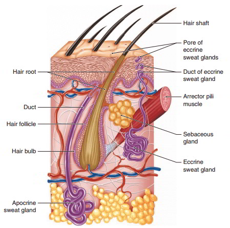
Epidermis And Accessory Structure Formed By The Epidermis And Their Functions
Structure of the Heart | The Franklin Institute The heart consists of four chambers: two atria on the top and two ventricles on the bottom. Looking at the Valentine’s Day heart, the two rounded humps at the top are rounded like the top of a lower-case “a.” The bottom is shaped like a “v.” Feel it working What else is inside your heart?

The Heart Diagrams Labeled and Unlabeled
Labelling the heart — Science Learning Hub Blood transports oxygen and nutrients to the body. It is also involved in the removal of metabolic wastes. In this interactive, you can label parts of the human heart. Drag and drop the text labels onto the boxes next to the diagram. Selecting or hovering over a box will highlight each area in the diagram.

In this diagram they are showing the function of the heart as they have labels to the parts of ...
Human Heart Models | Heart Anatomy Models | Vitality Medical The heart model with labels is hand-painted with vivid colors to illustrate the papillary muscles, heart valves, and adjacent structures. Sort By 4 Items Magnetic Heart Model, Life Size, 5 Parts $327.45 View Details Human Heart Model $450.66 - $566.36 View Details Classic Heart Model $81.03 View Details Magnetic Heart Model, Life Size, 5 Part G01
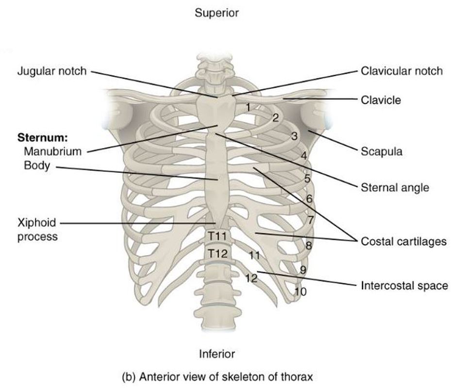
the Thoracic Cage - SCIENTIST CINDY
Label Heart Anatomy Diagram Printout - EnchantedLearning.com Every day, the heart pumps about 2,000 gallons (7,600 liters) of blood, beating about 100,000 times. Label the heart anatomy diagram below using the heart glossary. Note: On the diagram, the right side of the heart appears on the left side of the picture (and vice versa) because you are looking at the heart from the front. Enchanted Learning Search

MRI BLOG: Cardiac Anatomy
Heart Anatomy: Labeled Diagram, Structures, Blood Flow ... The first 2 structures are responsible for carrying deoxygenated blood from the body to the right side of the heart (right atrium). They are known as the superior vena cava and inferior vena cava. Their names are easy to remember because we know blood vessels that carry blood to the heart are veins, and this will help you think of vena cava.

Kids' Health - Topics - Your heart | Heart for kids, Heart diagram, Heart lesson
Label the heart — Science Learning Hub Label the heart Interactive Add to collection In this interactive, you can label parts of the human heart. Drag and drop the text labels onto the boxes next to the diagram. Selecting or hovering over a box will highlight each area in the diagram. Right ventricle Right atrium Left atrium Pulmonary artery Left ventricle Pulmonary vein Semilunar valve
Anatomy Of The Heart Quiz Label
Structure of the Heart | SEER Training Structure of the Heart The human heart is a four-chambered muscular organ, shaped and sized roughly like a man's closed fist with two-thirds of the mass to the left of midline. The heart is enclosed in a pericardial sac that is lined with the parietal layers of a serous membrane. The visceral layer of the serous membrane forms the epicardium.
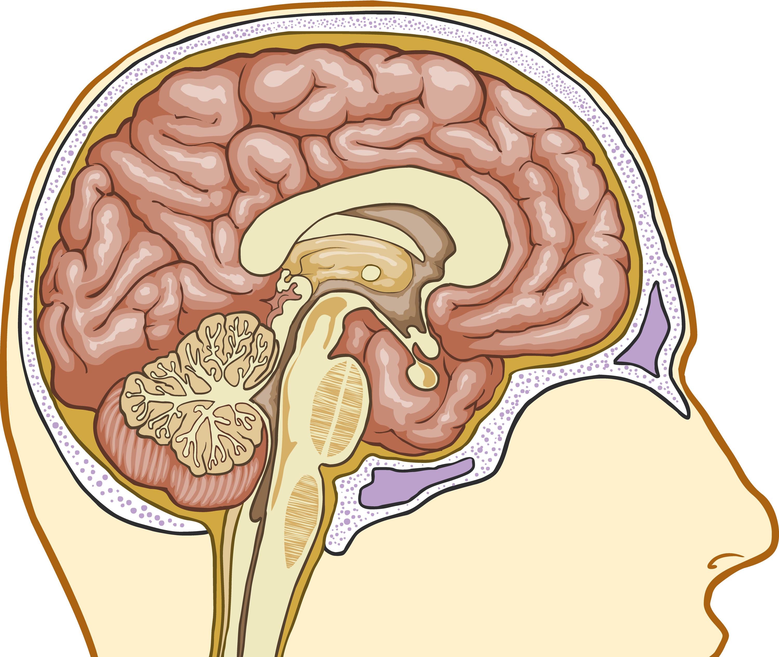
Basic Structure Of The Thalamus - Interactive Biology, with Leslie Samuel
Human Heart - Anatomy, Functions and Facts about Heart Following are the main functions of the heart: One of the primary functions of the human heart is to pump blood throughout the body. Blood delivers oxygen, hormones, glucose and other components to various parts of the body, including the human heart. The heart also ensures that adequate blood pressure is maintained in the body.
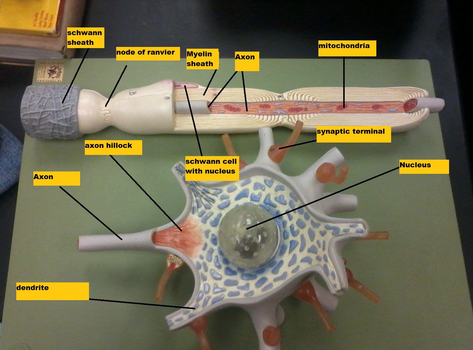
Chapter 14 Nervous Tissue - Biology 4 Human AnatomyProfessor Julie GallagherBarstow Community ...
Human Heart - Diagram and Anatomy of the Heart - Innerbody Because the heart points to the left, about 2/3 of the heart's mass is found on the left side of the body and the other 1/3 is on the right. Anatomy of the Heart Pericardium. The heart sits within a fluid-filled cavity called the pericardial cavity. The walls and lining of the pericardial cavity are a special membrane known as the pericardium.

Please label the following heart anatomy: Quiz
Heart anatomy: Structure, valves, coronary vessels | Kenhub The heart is shaped as a quadrangular pyramid, and orientated as if the pyramid has fallen onto one of its sides so that its base faces the posterior thoracic wall, and its apex is pointed toward the anterior thoracic wall.

Heart label diagram
Related Items
Post a Comment for "41 heart structure and labels"