43 human heart diagram without labels
A pan-tissue DNA methylation atlas enables in silico ... Mar 11, 2022 · a, Diagram illustrating the 13 tissue types chosen to build the DNAm-atlas resource, a database of corresponding tissue-specific DNAm reference matrices.Flowchart depicts the construction of a ... Heart Labeling Quiz: How Much You Know About Heart ... Create your own Quiz Here is a Heart labeling quiz for you. The human heart is a vital organ for every human. The more healthy your heart is, the longer the chances you have of surviving, so you better take care of it. Take the following quiz to know how much you know about your heart. Questions and Answers 1. What is #1? 2. What is #2? 3.
Dopamine - Wikipedia Structure. A dopamine molecule consists of a catechol structure (a benzene ring with two hydroxyl side groups) with one amine group attached via an ethyl chain. As such, dopamine is the simplest possible catecholamine, a family that also includes the neurotransmitters norepinephrine and epinephrine.

Human heart diagram without labels
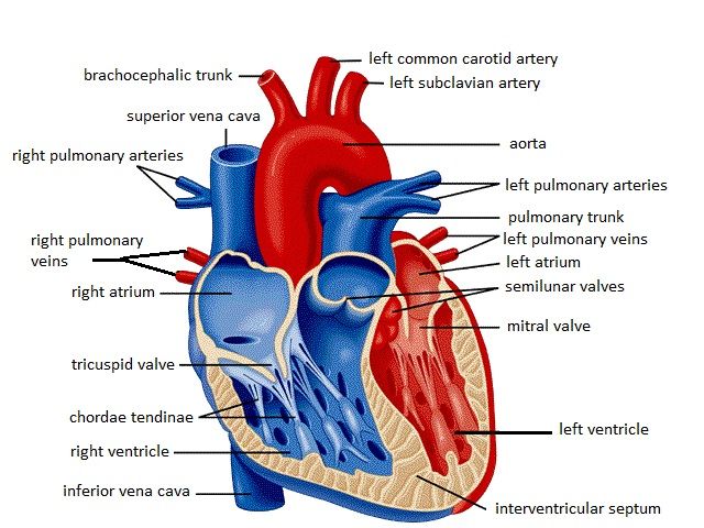
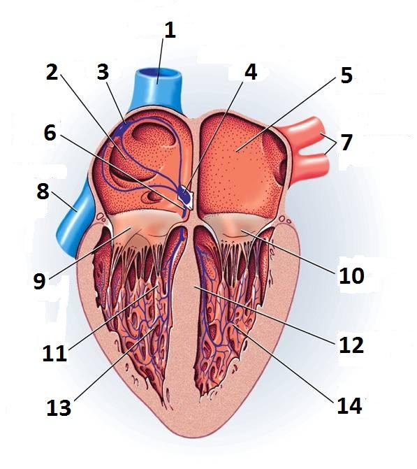
Heart Anatomy: Labeled Diagram, Structures, Blood Flow ... Image: Cardiac anatomy diagram showing the right and left side of the heart. The right side includes chambers 1 and 2. The left side includes chambers 3 and 4. Top vs Bottom of the Heart Next, we can divide the top 2 chambers of the heart from the bottom 2 chambers. The 2 chambers on top are known as the atria, and they include boxes 1 and 3. How to Draw a Human Heart: 11 Steps (with Pictures) - wikiHow The heart works like a pump and beats 100,000 times a day. The heart has two sides, separated by an inner wall called the septum. The right side of the heart pumps blood to the lungs to pick up oxygen. The left side of the heart receives the oxygen-rich blood from the lungs and pumps it to the body. The Anatomy of the Heart - Quiz 1 - Free Anatomy Quiz The circulatory system - lower body image, with blank labels attached. The circulatory system - a PDF file of the upper and lower body for printing out to use off-line. Describe and explain the function of the circulatory system - The circulatory system consists of the heart, the blood vessels (veins, arteries, and capillaries), and the blood.
Human heart diagram without labels. Human Heart - Diagram and Anatomy of the Heart The heart is a muscular organ about the size of a closed fist that functions as the body's circulatory pump. It takes in deoxygenated blood through the veins and delivers it to the lungs for oxygenation before pumping it into the various arteries (which provide oxygen and nutrients to body tissues by transporting the blood throughout the body). Diagram of Blood Flow Through the Heart - Bodytomy You must have seen the human heart diagram. Thus, you will be aware of the anatomy of the human heart. If not, you can have a look at the labeled diagram of the human heart present in this article. The strongest muscle in the human body is the human heart. The human heart continues to pumps liters of blood throughout the body all lifelong. Organ Map | Diagram of Human Body Internal Organs Functions Featuring an accurate illustration and informative, clear labels, this organ map is a fantastic visual aid to support your teaching during science lessons all about the human body's internal organs.Once downloaded, you'll have an A4 diagram of a human body and internal organs, clearly labelled and perfect for individual use. Each internal organ is labelled and includes a definition of each ... Human Heart Diagram Without Labels | Human heart diagram ... ABOUT THIS ACTIVITY: Illustrates the pathway of blood through the heart. The areas of the heart with MORE oxygen are labeled with an "R". Students will color these areas RED. The areas of the heart with LESS oxygen are labeled with a "B". Students will color these areas BLUE.
Explain Block Diagram of Computer and Its Components Apr 14, 2022 · The above block diagram of Computer represents the complete process of an internal computing system. Central Processing Unit (Cpu) The abbreviation of CPU is Central Processing Unit. It is generally defined as brain and heart of any computer System. It comprises of three main components such as Memory Unit, Control Unit, Arithmetic and Logic ... Human Heart Diagram Labeled - Science Trends List Of Heart Structures Heart Chambers Ventricles - The bottom two heart chambers. Atra - The upper two heart chambers. Wall Of The Heart Sinoatrial Node - A collection of tissue that releases electrical impulses and defines the rate of contraction for the heart. Atrioventricular Bundle - The fibers which transmit cardiac impulses. Human heart diagram Images, Stock Photos & Vectors ... Human heart diagram royalty-free images. 14,543 human heart diagram stock photos, vectors, and illustrations are available royalty-free. See human heart diagram stock video clips. Image type. How to Draw the Internal Structure of the Heart (with ... To draw the internal structure of a human heart, follow the steps below. Part 1 Finding a Diagram 1 To find a good diagram, go to Google Images, and type in "The Internal Structure of the Human Heart". Find an image that displays the entire heart, and click on it to enlarge it. 2 Find a piece of paper and something to draw with.
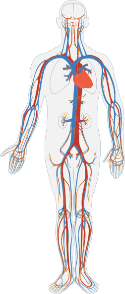
Human Heart Images | Free Vectors, Stock Photos & PSD Human internal organs anatomy in cartoon vector style. brain and kidney, liver and lung, stomach and heart illustration. macrovector. 104. Like. Collect. Save. Cute heart set. human organ with face and different emotions, happy, sad, angry, sick and healthy cartoon character. v. pch.vector. Circulatory System Diagram - Cardiovascular System and ... They may come with or without labels. Common circulatory system diagrams show pulmonary circulation, coronary circulation, systematic circulation, veins, arteries, or a combination. The systemic circulation system is the most commonly illustrated of the systems that make up the circulatory system as it is the largest. Human Heart - Anatomy, Functions and Facts about Heart The human heart is one of the most important organs responsible for sustaining life. It is a muscular organ with four chambers. The size of the heart is the size of about a clenched fist. The human heart functions throughout a person's lifespan and is one of the most robust and hardest working muscles in the human body. (PDF) Heart Disease Prediction System - ResearchGate Globally, cardiovascular (heart) diseases are the major cause of death. About 80% of deaths are reported in developing countries. Looking at the trend and lifestyle, one can predict that by 2030 ...
a Lebelled Diagram of the Heart : Anatomy and physiology Heart - Cancer / Asbestos Cancer ...
Heart Blood Flow | Simple Anatomy Diagram, Cardiac ... Step 1 involves blood vessels, similar to what we saw with step 1 in the right side of the heart. The pulmonary veins carry oxygenated blood from the lungs to the left side of the heart, specifically the left atrium. There will be better images of the pulmonary veins shown in the images later in this post. 2. Left Atrium

1000+ images about School on Pinterest | Heart diagram, Physician assistant and Medical
circulatory system worksheet without labels - Google ... Nov 3, 2015 - circulatory system worksheet without labels - Google Search. Nov 3, 2015 - circulatory system worksheet without labels - Google Search. Pinterest. Today. ... Human Heart Diagram. Anatomy Coloring Book. Coloring Sheets. Colouring. Heart Coloring Pages. Heart Anatomy. More information...
Unlabelled Diagram Of The Heart - Cliparts.co
Venn Diagram in R (8 Examples) | Single, Pairwise, Tripple ... We should usually do this step before the creation of each venn diagram, because otherwise the venn diagram is just overlaying previously created plots. Second, we are producing our single venn diagram with the draw.single.venn function. All we are specifying within the function is the size of our area (i.e. 10). Example 2: Pairwise Venn Diagram

u414adad: heart diagram without labels
A Labeled Diagram of the Human Heart You Really Need to ... The human heart, comprises four chambers: right atrium, left atrium, right ventricle and left ventricle. The two upper chambers are called the left and the right atria, and the two lower chambers are known as the left and the right ventricles. The two atria and ventricles are separated from each other by a muscle wall called 'septum'.

Heart Diagram stock vector. Illustration of vector, cardiovascular - 42694065
Human Heart (Anatomy): Diagram, Function, Chambers ... The heart is a muscular organ about the size of a fist, located just behind and slightly left of the breastbone. The heart pumps blood through the network of arteries and veins called the...
Biology : Human Organ Systems III - Worksheet / Test Paper
Heart Diagram Unlabeled - Cliparts.co 84 images of Heart Diagram Unlabeled. You can use these free cliparts for your documents, web sites, art projects or presentations. Don't forget to link to this page for attribution!

Heart Diagram Drawing at GetDrawings.com | Free for personal use Heart Diagram Drawing of your ...
Anatomy of a Human Heart - uofmhealth Parts of the human heart . The heart is made up of four chambers: two upper chambers known as the left atrium and right atrium and two lower chambers called the left and right ventricles.. MORE FROM MICHIGAN: Sign up for our weekly newsletter. It is also made up of four valves: the tricuspid, pulmonary, mitral and aortic valves.

STUDY GUIDE
13+ Heart Diagram Templates - Sample, Example, Format ... cfep.uci.edu This response sheet of blood and the heart contains all the parts of human heart including arteries, veins, atrium and so on. All the minute parts inside the human heart have been clearly labelled. Free Download Heart Structure And Its Functions Pdf Format
The Heart: Structure and Function - YouTube
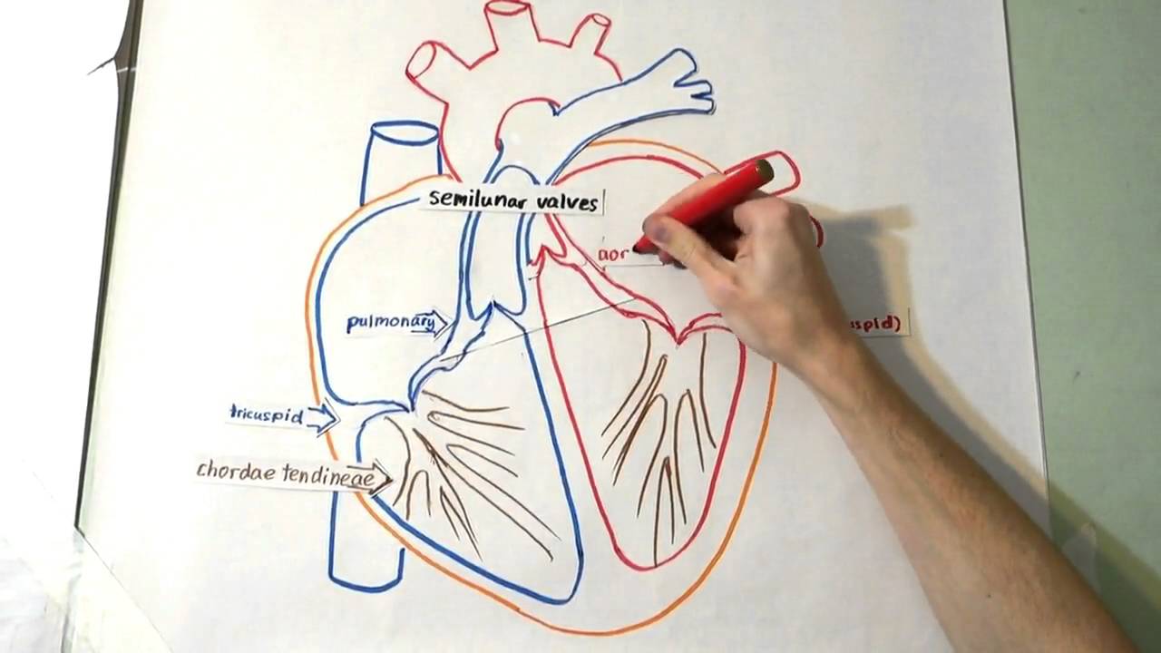
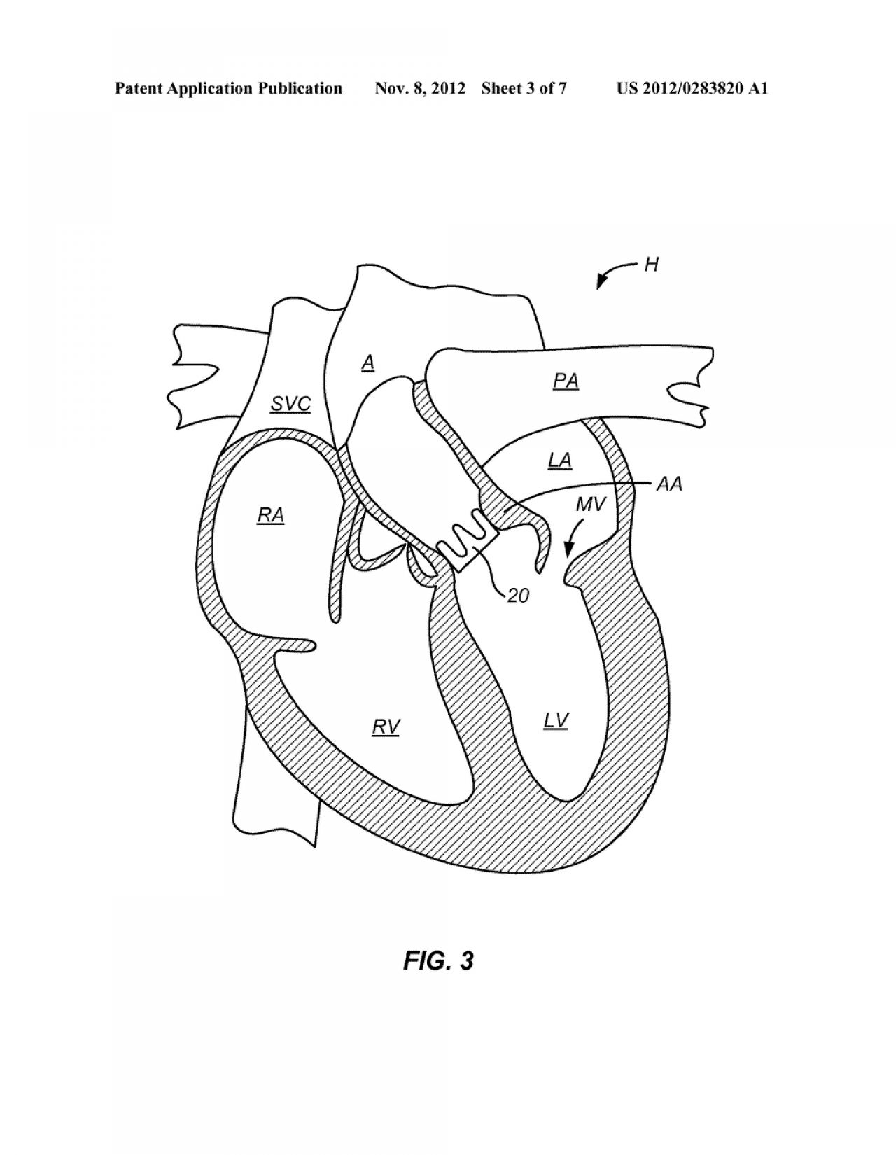
PDF Anatomy of Heart Labeled and Unlabeled Images (a) Anterior view of the external heart C' 2019 Pearson Education. Aort'c arch Ligamentum arteriosum Left pulmonary artery Left pulmonary ve ns Auricle of left atrium Circumflex artery Left coronary artery (in atrioventricular sulcus) Great cardiac vein Left ventricle Anterior interventricular artery (in anterior interventricular sulcus) Apex
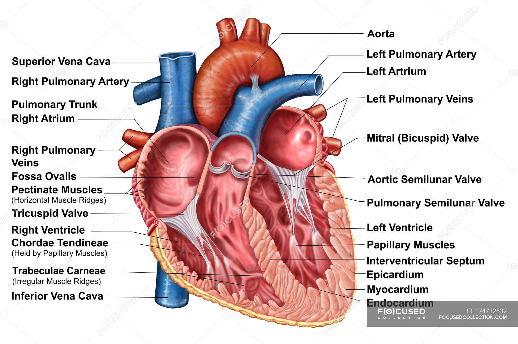
Anatomy of heart interior with labels — cross section, blood vessels - Stock Photo | #174712532
Heart Diagram - 15+ Free Printable Word, Excel, EPS, PSD ... Heart Diagram - 15+ Free Printable Word, Excel, EPS, PSD Template Download A heart diagram is a popular design used by different people for various uses. It can be used by a teacher or student for academic purpose, by a friend or relative for mutually sending and exchanging cards or for baby toys or printing on dresses etc.
Unlabeled Heart Diagram - Cliparts.co
Heart Diagram with Labels and Detailed Explanation - BYJUS The human heart is the most crucial organ of the human body. It pumps blood from the heart to different parts of the body and back to the heart. The most common heart attack symptoms or warning signs are chest pain, breathlessness, nausea, sweating etc. The diagram of heart is beneficial for Class 10 and 12 and is frequently asked in the ...
Free Blank Heart Diagram, Download Free Blank Heart Diagram png images, Free ClipArts on Clipart ...
Label the heart - Science Learning Hub In this interactive, you can label parts of the human heart. Drag and drop the text labels onto the boxes next to the diagram. Selecting or hovering over a box will highlight each area in the diagram. Pulmonary vein Right atrium Semilunar valve Left ventricle Vena cava Right ventricle Pulmonary artery Aorta Left atrium Download Exercise Tweet

Emma Woolley...: Anatomical Diagrams
Anatomy Chart - How to Make Medical Drawings ... - SmartDraw Anatomy Chart What is an Anatomy Chart? An anatomy chart refers to a visual depiction of the human body. It can show the entire body or focus on a particular system using systemic anatomy such as the muscular, skeletal, circulatory, digestive, endocrine, nervous, respiratory, urinary, reproductive, and other systems. There are many different branches of anatomy dealing with the human body ...
Circulatory System No Labels Clip Art at Clker.com - vector clip art online, royalty free ...
Human Heart Diagram Without Labels - Labelling Worksheet There are two versions of this resource. The standard version comes with two pages - the first has a human heart diagram without labels, and the second has the answers. There's also a version for pupils working at a lower ability, which has the same human heart diagram without labels, and the answers in a box underneath for pupils to consult.

34 Label Of Heart Diagram - Labels Database 2020
Geography News -- ScienceDaily Geography. Read the latest geographical research from universities and institutes around the world.
Post a Comment for "43 human heart diagram without labels"