40 basic animal cell diagram with labels
Animal Cell - Structure, Function, Diagram and Types The most common types of animal cells are: Skin Cells Melanocytes, keratinocytes, Merkel cells and Langerhans cells Muscle Cells Myocyte, Myosatellite cells, Tendon cells, Cardiac muscle cells Blood Cells Leukocytes, erythrocytes, platelet Nerve Cells Schwann cell, glial cells etc Fat Cells Adipocytes Points to Note About Animal Cell Free Animal Cell Diagram Templates - Edrawsoft Animal Cell Diagram Template. A clear design animal cell diagram template from Edraw is waiting for you in the free download version. Use it for any kinds of science coursework or group discussions. You can also adjust the diagram sizes at any time you want for more insights. Lab Apparatus List. 64703.
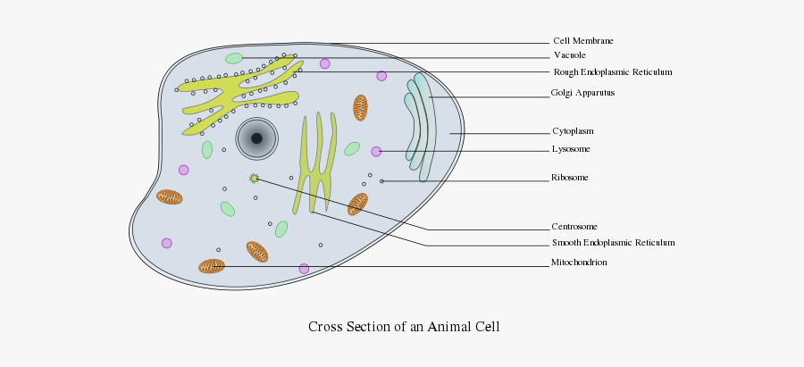
Animal Cell Diagram | Science Trends An animal cell diagram is a great way to learn and understand the many functions of an animal cell. The diagram, like the one above, will include labels of the major parts of an animal cell including the cell membrane, nucleus, ribosomes, mitochondria, vesicles, and cytosol.

Basic animal cell diagram with labels
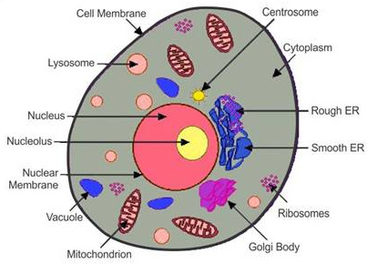
Label the Animal Cell - 4th Grade Science Worksheet - SoD Label the Animal Cell Label the Animal Cell. The basic functional and structural unit of all living organisms, including animals, consists of cells. Cells are complex entities that keep changing their form and blending with other cells. This free printable 4th grade science worksheet helps you learn more about the structure and parts of animal ... Animal Cells: Labelled Diagram, Definitions, and Structure Animal Cells Organelles and Functions. A double layer that supports and protects the cell. Allows materials in and out. The control center of the cell. Nucleus contains majority of cell's the DNA. Popularly known as the "Powerhouse". Breaks down food to produce energy in the form of ATP. › mmwr › previewGuidelines for Safe Work Practices in Human and Animal ... Jan 06, 2012 · The guidelines in this section are combined biosafety best practices for both human autopsy and human surgical pathology and animal necropsy and veterinary surgical pathology. When necessary, biosafety guidelines specific for human or animal diagnostic laboratory settings are highlighted. 5.1. Autopsy/Necropsy–Associated Infections
Basic animal cell diagram with labels. A Well-labelled Diagram Of Animal Cell With Explanation Well-Labelled Diagram of Animal Cell The Cell Organelles are membrane-bound, present within the cells. There are various organelles present within the cell and are classified into three categories based on the presence or absence of membrane. Listed below are the Cell Organelles of an animal cell along with their functions. Animal cells - Cell structure - AQA - GCSE Combined ... Animal cells have a basic structure. Below the basic structure is shown in the same animal cell, on the left viewed with the light microscope, and on the right with the transmission electron... A Labeled Diagram of the Animal Cell and its Organelles ... One can observe the golgi apparatus in the labeled animal cell parts diagram. The golgi apparatus is situated near the cell nucleus and besides the stacked sacs, it also contains large number of vesicles. The main function of this golgi complex is to receive the proteins synthesized in the ER and transform it into more complex proteins. Blank Animal Cell Diagram To Label Simple : Functions and ... File:Simple diagram of animal cell (blank).svg - Wikimedia … (Albert Briggs) We have a great hope these Animal Cell Labeling Worksheet images collection can be useful for you, bring you more ideas and also help you get an amazing day. RER (Rough Endoplasmic Reticulum) synthesizes proteins.
› programs › science-netlinksScience NetLinks | American Association for the Advancement ... Mar 09, 2022 · Our ability to provide a voice for scientists and engineers and to advance science depends on the support from individuals like you. Give Animal And Plant Cell Diagram To Label Worksheets ... 12. $2.00. PDF. Three versions of the plant cell worksheet and three versions of the animal cell worksheet allow students of different grade levels and/or skill levels to label and review the parts of each cell type. Can be used as homework or a quiz. Answer key is provided and can be projected and used during less. Complete Guide! Animal Cell Diagram, Parts of an Animal ... The organelles found in most animal cells include the nucleus, cell membrane, cytoplasm, mitochondria, ribosomes, lysosomes, vacuoles, centrosome, endoplasmic reticulum, and Golgi apparatus. Let's discuss these organelles in detail: Cell Membrane This is the outer barrier of the animal cell. It controls what goes in and out of a cell. Animal and Plant Cell Worksheets Download and print worksheets for teaching students about animal and plant cells. We have cell diagrams with and without labels, as well as vocabulary activities. Animal Cells (Basic) Identify Animal Cell Parts Write the name of each animal cell part shown in the diagram. The illustration includes definitions. View PDF Filing Cabinet
Label the Animal Cell - EnchantedLearning.com Golgi body - (also called the Golgi apparatus or Golgi complex) a flattened, layered, sac-like organelle that looks like a stack of pancakes and is located near the nucleus. It produces the membranes that surround the lysosomes. The Golgi body packages proteins and carbohydrates into membrane-bound vesicles for "export" from the cell. › applications › fretBasics of FRET Microscopy | Nikon’s MicroscopyU The search for an ideal red-emitting fluorescent protein has long been the goal for live-cell and whole animal imaging using FRET biosensors and fusions, primarily due to the requirement for probes in this spectral region in multicolor imaging experiments as well as the fact that longer excitation wavelengths generate less phototoxicity and can ... en.wikipedia.org › Cell_membrane_(diagrammatic)Wikipedia:Featured picture candidates/Cell membrane ... Image:Plant_cell_structure_svg.svg, a Featured Picture, is under the same threat of summary deletion, as are many of LadyofHats (Mariana Ruiz) other contributions, for example Image:Human arm bones diagram.svg a FP, Image:Average prokaryote cell- en.svg, a FP, Image:Animal cell structure.svg, the FPC below... Animal Cell Diagram Labeled | EdrawMax Template According to the animal cell labeled diagram, some of the cell organelles of an animal cell are the cell membrane, Cytosol, Cytoskeleton, nucleus, Ribosomes, Endoplasmic Membrane, Vesicles, Mitochondria, and more. Creating such labeled diagrams will teach your young students more about animal cells.
41 best images about BEI Science Year 5 - Cells on Pinterest | Cell structure, Of life and Life ...
Animal Cell- Definition, Structure, Parts, Functions ... The largest animal cell is the ostrich egg which has a 5-inch diameter, weighing about 1.2-1.4 kg and the smallest animal cells are neurons of about 100 microns in diameter. Animal cells are smaller than the plant cells and they are generally irregular in shape taking various forms of shapes, due to lack of the cell wall.
Animal Cell Diagram Labeled Gcse / Biology B2 | Biology, Science classroom, Classroom displays ...
Plant and Animal Cell: Labeled Diagram, Structure ... Both plant and animal cells have similar types of architecture. They are made up of cell boundaries, cytoplasm, nucleus and several cellular organelles. Structure. Description and function. Cell Wall. 1. Non-living, rigid, outer boundary. 2. Made up of cellulose, hemicellulose, pectin, lignin, etc.

The cell is like a hotel
Animal Cell Diagram To Label Simple : Functions and Diagram A worksheet with a simple diagram to label the main subcellular structures (Nucleus, Mitochondria, Ribosomes, Cell membrane and Cytoplasm) of an Animal cell. It's the cell's brain, employing chromosomes to instruct other parts of the cell. Anatomynote.com found Animal Cell Diagram Label from plenty of anatomical pictures on the internet.
Animal Cells Labeled Diagram ~ DIAGRAM
Well Labelled Diagram of Animal Cell - Detailed ... The different organelles shown in a well labelled diagram of an animal cell are explained below: Cell Membrane/Plasma Membrane To draw a well labelled diagram of an animal cell, the cell membrane has to be drawn. The cell membrane is an integral part of the cell structure that keeps the entire cell bound together.
What cells don’t take part in cell division? - Quora
› category › newsNews Archives | Hollywood.com Travel through time by exploring Hollywood.com's entertainment news archives, with 30+ years of entertainment news content.

Grade 9 Animal Cell Diagram
Labeled Animal Cell Diagram with Definitions Labeled Animal Cell Diagram with Definitions Posted 8 years ago 4800 Parts and their Functions Cell Membrane: The cell membrane is the outer most part of the cell which encloses all the other cell organelles. The cell membrane controls the influx of the nutrients and minerals in and out of the cell.

Animal Cell
Animal Cell Diagram with Label and Explanation: Cell ... Animal cell is a typical Eukaryotic cell enclosed by a plasma membrane containing nucleus and organelles which lack cell walls, unlike all other Eukaryotic cells. The typical cell ranges in size between 1-100 micrometers. The lack of cell walls enabled the animal cells to develop a greater diversity of cell types.

Human Behavior and the Social Environment I BSW: Cell Diagram 2
Diagram Of Plant And Animal Cells To Label Teaching ... Google Drive™ folder. This worksheet includes both an extensive animal and plant cell diagram to label. The animal cell includes 17 organelles, and the plant cell includes 20 organelles for students to label and color. There is also a 4 page graphic organizer (chart) that includes a drawing of each of the organelles in alphabetical order.
Post a Comment for "40 basic animal cell diagram with labels"